![]() A picture is worth a thousand words. Take a picture, it’ll last longer.
A picture is worth a thousand words. Take a picture, it’ll last longer.
Picture this:
 You are visiting the beautiful Oregon Coast. The sun is shining. The breeze is whispering softly through the dune grass. A gust blows the foam back from the tops of the waves. The water sparkles like diamonds in the afternoon sun. The sun-warmed sand massages your feet as you wander along the water’s edge, dancing just out of reach of the cold northern Pacific waves. The contrast between sand, air, and water temperatures is startling. As the sun descends toward the edge of the horizon, you stop, gazing out across the vast deep. The azure blue sky slowly changes. oranges, reds, yellows, purples, pinks, even green, and then darker blue. Darkening ever so slightly as the sun kisses the western horizon, and then slowly sinks beneath the waves. The sky continues to darken. Suddenly, as if by magic, pinpoints of light appear. They seem to shimmer and sparkle, much like the diamond waves. In the east, the grand white of the moon ascends, grinning for all the world to see like the mythic Chestershire cat.
That word picture is just under 200 words. I’m sure I could add more detail, but I am afraid if I were to do that, I would abandon my post and run away to the coast, without a backward glance. However, if you look at the photos I found you would see instantly what I was describing, and in much less space and time. And what if you have never been to the Oregon Coast? Would my description give you an accurate representation? Or is it only by your experience, combined with my words, that gives you the sense of peace (and longing) that comes with visiting the ocean, sitting for hours as the waves roll and the sun moves through the sky?
Often in medicine, human and veterinary (and, I have heard, even in botany), doctors turn to imaging to assist in diagnosing injury and illness. Would that we could all be like Superman, with his x-ray vision. Then we could simply  gaze within to understand what ails. Alas, we are not so lucky as he. Fortunately though, we live in a time that is replete with advances in medical imaging and diagnostics. Each is similar in its function, and yet each has a specific use and application. Recently, I heard several terms used that I was unfamiliar with. Being the inquisitive cat that I am, I decided to check them out. I also wondered how many others, like me, might be out there. If you understand what some of the different imaging options are, it might help you out should you ever be in a situation that would require additional diagnostic tools.
I spoke with my new wonderful friend Dr Nile about the different types of imaging. She also directed me to some websites on the internet. My thanks to the folks at www.medicinenet.com, UC Davis School of Veterinary Medicine, www.radiologyinfo.org, and Wikipedia, for helping me with understanding the medical jargon. Some of those websites out there are really technical. While I consider myself quite evolved, I also have come to realize there is a reason I am a cat and not a doctor. (Though I would love to play one on TV). I compiled a list of the different modes of medical imaging for your information.
 X-RAY is one of the oldest and perhaps best known forms of medical imaging, dating back to Germany in 1895. Radiation is formed into a beam that passes through objects at different rates, depending on their density (thickness). It strikes a photographic surface to produce an image, of teeth, bones, or organs, called a radiograph. Doctors can then look at the radiograph to assess the overall size, shape, number, density (amount of grayness in the color of the image) and location of different organs as well as locate any foreign material that should not be there. Examples of foreign material would include perhaps a rock or ball in a dog’s stomach, or a BB bullet in a cat’s side. Radiographs are available in most, if not all, general practice offices. Depending on the presenting problems and symptoms, radiographs are often one of the first diagnostic tools recommended for pets after a thorough physical exam.
 Ultrasound (sonogram, echocardiogram) uses sound waves to create images. The sound waves are higher than those audible to humans and echo off the tissues of the body. Different tissues reflect varying degrees of sound waves and are recorded as images by the ultrasonographer. Ultrasound (or sonography) is most useful in viewing soft tissues. A hand held probe called a transducer is moved over an area of the body to image the structures below. It is often used in pregnancy to visualize the fetus and is usually the most well-known use of ultrasound technology. However, it can be used in virtually all areas of the body, to varying degrees. Cardiac ultrasound (echocardiogram) helps doctors visualize the function of the heart valves and
ventricles. In abdominal sonography, the solid organs of the abdomen such as liver, kidneys, aorta, gall bladder, and spleen are imaged. Sound waves can be blocked by gas and weakened by fat, so the capabilities can be limited but still useful. Ultrasound can be used to image the lungs, eyes, the urinary systems, as well as tendons and muscles. It can even be used as an alternative to x-rays in very young patients, if medically indicated.
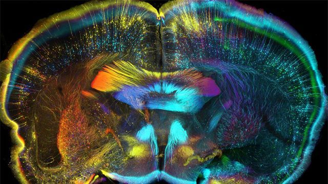
 MRI, or Magnetic Resonance Imaging, uses a strong magnet to produce and radio wave energy to produce images of organs and internal structures. It is used by doctors to identify injury, bleeding, tumor, infection, or other injury. Doctors can diagnose and monitor brain and spinal injuries and disorders, joint disorders or abnormalities, organ tumors or abnormalities, and assess the size and function of the heart. It does not use radiation in its imaging, and is a specialty service available at some hospitals.
CT is an abbreviation for Computed (or computerized) Tomography. (Tomography refers to imaging by sections through the use of a penetrating wave.) It is essentially a series of x-rays taken in “slices” through the body. These are then analyzed by a computer and reconstructed as 3D or 4D image. CT can help to diagnose muscle or bone disorders, identify the location of infections, tumors or blood clots, and detect internal injury. CT images are extremely useful in guiding surgical or biopsy procedures and help in monitoring certain conditions like cancer. CT scans are often a referral procedure and are performed at specialty hospitals, as is the PET scan.
PET scan stands for Positron Emission Tomography. Like many of the imaging tests, it uses radioactive positrons (positively charged particles) to detect differences in chemical and metabolic activity within the body. An area with increased activity shows on a colored image. This type of scanning shows function of body systems, rather than looking at or for structures as in other types of imaging. PET scans are very useful in looking for cancer activity which generally shows as more metabolic activity, and can also detect very small areas of activity. They can also differentiate areas of scar tissue, which tend to have very low rates of metabolic activity.
I nterventional Radiology provides minimally invasive image-guided diagnosis and treatment of disease in every organ system. It is also called surgical radiology. The range or procedures performed by interventional radiologists is broad, but the unifying concept behind it is to provide the most modern and least invasive technique available for diagnosis and treatment, so as to minimize risk to the patient and improve medical outcome. Methods of interventional radiology include x-ray, CT, ultrasound, and MRI. Procedures are less invasive and recovery time is quicker. Hospital stays are shorter and infection rates are decreased. Examples would include target radiation treatment for cancer, or ultrasound-guided sample collection such as urine, or tumor biopsy.
 Fluroscopy is a technique of medical imaging which uses x-rays to obtain real-time moving images. A fluoroscope allows the doctor to view the internal structures and function of the patient. The pumping action of the heart or the motion of swallowing can be watched. It is similar to CT in that it produces images with x-rays, but differs in that it generates moving pictures so the action of a particular structure can be observed. Fluroscopy is performed by a radiologist and is typically available at a specialty hospital or university.
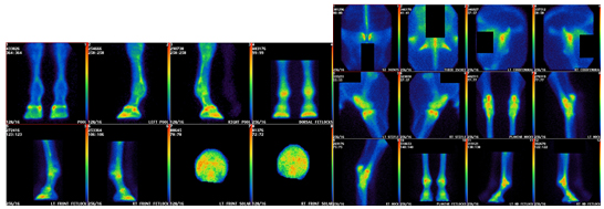 Scintigraphy comes from the Latin for “spark” and is a test used in nuclear medicine. Radioisotopes are taken internally and the emitted radiation is captured by specialized cameras that detect gamma radiation and form two-dimensional images called projections. Scintigraphy has application in diagnosing various disease processes in almost every body system, from the heart and lungs, to the liver, kidney, and biliary systems. As with several of the different medical imaging techniques, scintigraphy is a specialty procedure that requires advanced training in the safe use of radiation.
 Medical Photography is a specialized area of photography that documents the clinical presentation of patients, medical and surgical procedures, and medical devices. Medical photographers document patients at various stages of illness, before and after surgical procedures, and various stages of injury. Medical photographs are used as part of the medical record of a patient as well as training and education of medical professionals and the public. Rest assured, when Dr Nile pulls out her phone to take a picture of your pet, it is to help her track your pet’s recovery process.
X-ray, ultrasound, and medical photography are diagnostic procedures that can usually be performed in your veterinarian’s general practice office. The more specialized imaging options are performed at veterinary hospitals with radiologists on staff. Radiologists are doctors of veterinary medicine that have gone received specialized training and become certified in medical imaging and the safe use of radiation. If you would like to learn more about radiologists and what they do, visit www.avcr.org/page/pet-owners.
Lots of options, lots of applications. All with the goal of better diagnostics and healthier, longer lives.
Oh, and by the way, the way the clouds played over Mt. McLoughlin the other day? No words could describe it, so here’s a picture. Hope you enjoy it as much as I do!