SAN DIEGO—As I stepped into the Brain Observatory, I didn’t really see what I expected from a brain bank. Sure, there’s a glassed-in lab space and a refrigerator with a few Tupperware-clad brains. But the walls are bedecked with art, and the sleek modern sofas give off more of a nightclub feel than a sterile lab vibe.
Coincidently, this isn’t a traditional brain bank. Instead of specimens lined up on shelves and stacked in massive freezers, neuroanatomist Dr. Jacopo Annese is curating a vast virtual collection of brains that will be housed online. Called the Digital Brain Library, it’s a major shift in how we store and study information gleaned from the brain.
Anatomists have been collecting and preserving humans’ soft tissue for more than 300 years; the practice stemmed both from an aspiration to understand our how our bodies work and a much seedier desire to sell organs on the black market.
Thanks to its jelly-like texture, the brain has always been one of the most difficult organs to work with. But through trial and error, scientists eventually developed ways to toughen and preserve the brain in order to allow it to be examined, dissected, and studied.
Most brain banks today function as repositories and distributors; once a brain is preserved, various parts and pieces are doled out to researchers with specific questions about a region or a disease. What’s left of the brain sits a freezer until it is requested. It’s a practical system but not a holistic one. All sense of connectivity—of how the brain’s regions work together as an entire organ—can easily get lost in the shuffle.
Connectivity is the holy grail of neuroanatomy, and it’s the guiding principle behind the Digital Brain Library. Instead of the traditional piece-by-piece approach to brain banking (which Annese jokingly refers to as “meat-packing”), the Brain Observatory aims to make entire brains accessible to anyone who may be interested.
Think of the project as the Google Earth of neuroscience.
A closer look
Annese defies the trope of the stuffy scientist; he looks like he might feel at home in the very same nightclub as his sofas. He strides in, just a little late, clad in a purple dress shirt and a crisp charcoal suit. He’s chatty and charming with a lilting Italian accent.
Currently, Annese’s lab is located in a low-slung, nondescript building at the University of California, San Diego. But he’s in the process of moving the Brain Observatory’s operations to the Institute for Brain and Society, the nonprofit he started in 2013.
After some introductory banter, we sat down at a computer to tinker with the Digital Brain Library, and it didn’t take long for even this totally untrained eye to catch onto its advantages.
For decades, magnetic resonance imaging has helped us look inside our own bodies, and today MRIs remain the best way to peek inside a living person’s head. But the painstaking—and proprietary—digitization process used at the Brain Observatory provides a totally unprecedented view of the brain, all the way down to its individual nuclei and axons.
“When you zoom in on an MRI image, really, at some point, you don’t know what the hell is going on in this voxel,” Annese demonstrated, pointing to a spot on an MRI. As he zoomed in, the image quickly dissolved into a blur.
“But when you really look into these voxels, this is what you find,” he continued, zooming in on a slice of brain that has been digitized for the Library. A beautiful, lacy blue image appears; nerve fibers that are just two or three microns wide are unmistakably sharp. For comparison, a human hair measures 75 microns or more. A single red blood cell is usually around five microns wide.
“When you talk about looking at structure in your brain, this is a level of resolution that corresponds to, like, single atoms for a physicist,” former Brain Observatory programmer Hauke Bartsch explained. “It’s very detailed, and we can look at structure in a way that is not possible if you just go into an MRI scanner.”
This high-res imagery has already drawn a host of users from the scientific and medical communities who are scouring the brain for treatments, preventions, and answers. But others are welcome to browse its collection as well; public access is a cornerstone of the Brain Observatory.
One benefit of this broad access is to raise the profile and awareness of the project; after all, brains are cool and science needs all the public support it can get. But allowing the public to comb through the Library also increases the likelihood that someone will unearth something fantastically interesting. Take Google Earth, for example, which amateurs have used to locate archaeological sites, discover North Korean secrets, and track the trajectories of meteorites. Citizen science projects have also allowed amateurs to find things professionals missed, such as an entire class of galaxies known as “green peas.” With enough curious eyes on a big enough dataset, you never know what someone will find.
Building a library
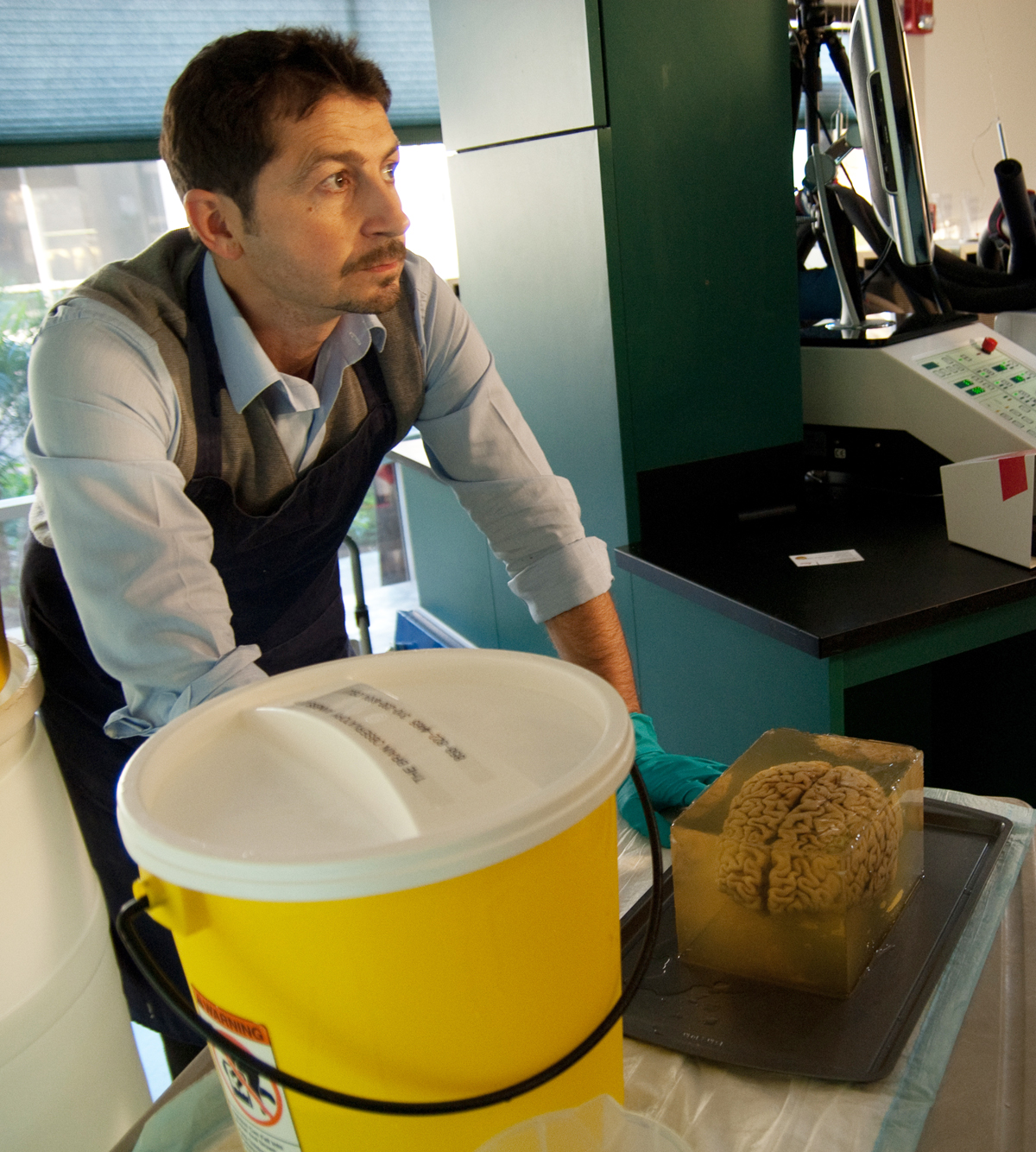
In order to tell us anything at all, each and every brain must be well-preserved. And that starts as soon as a donor dies, with an extended MRI scan once the family is ready to part with the body. Then, the brain is carefully removed from the head in a process called fenestration.
Initially, the brain is as soft as warm butter; it must be hung upside-down in formaldehyde in the lab’s refrigerator for several months to harden up the tissue. Then the brain takes another bath, this time in a sucrose solution. During this soak, the sugar displaces water molecules in the brain, better preserving the tissue when it’s frozen for slicing.
And slicing is the real marathon. A single brain produces more than 2,000 70-micron-thick slices in a painstaking process that can take days. The razor-sharp microtome blade cuts the brain automatically, but someone must be there to switch out the blade every 600 cuts or so and properly care for each of those priceless slices. In this video, you can watch Annese deftly use a paintbrush to swipe away each delicate section of H.M.’s brain, 2,401 slices in all. In another nod to open access, this entire 53-hour-long slicing session was live-streamed online.
“A lot of things can go wrong” during the slicing process, said lab assistant Colleen Sheh. “It’s manual, it’s a person that’s actually taking the slices. So it does take a lot of training before we let anyone sit and cut the brain.” And there’s a lot of pressure to get it right, since the project—not to mention the brains’ donors—have so much invested in each specimen.
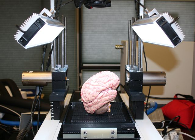
Once each slice is mounted, stains are applied to bring out the details of the brain’s structure (axons don’t normally have a blue hue like the ones I viewed). Then it’s on to digitization. One by one, each and every slice is photographed at 0.37 microns per pixel, a resolution 150 times smaller than the width of a human hair. At that fine a scale, it takes many stitched-together images to make up each slice; the brain’s largest sections—those that span from ear to ear—take more than 24 hours to photograph and digitize.
reader comments
65